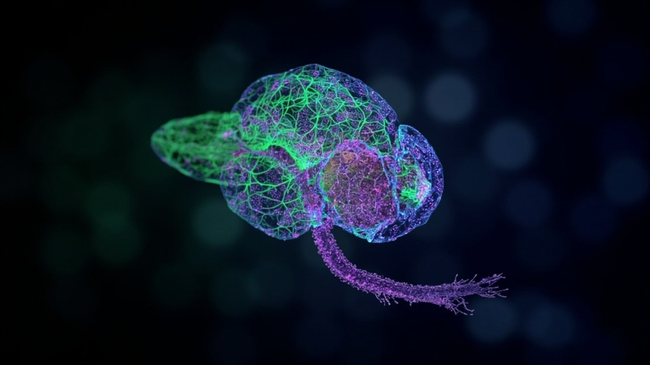
TL;DR: Scientists published the first complete wiring diagram of an adult male fruit fly's brain and nerve cord.
TL;DR: On September 3, 2026, a team led by HHMI Janelia Research Campus with Google Research and Cambridge collaborators published the complete connectome of the adult male fruit fly central nervous system, mapping more than 166,000 neurons and 125 million connections in Cell.
The release provides the first synaptic-resolution map of the entire male Drosophila melanogaster central nervous system, including the brain and ventral nerve cord, according to Google Research. The project reported over 166,000 neurons and 125 million synaptic connections, described as the largest brain map by number of neurons to date. The work was published in Cell under the title “Sexual dimorphism in the complete connectome of the Drosophila male central nervous system.”
The male map complements earlier female fruit fly brain maps and enables direct male-female comparison at synaptic resolution, according to Google Research. The Cell study reported 166,700 neurons spanning the brain and nerve cord, fully proofread and annotated, including 11,710 neuron types. Together with ongoing work on fish and mice, the authors described the wiring maps as a step toward revealing mechanics of how brains work.
A decade-long mapping project reaches completion
The male fruit fly connectome was the result of a decade-long partnership led by Howard Hughes Medical Institute’s Janelia Research Campus with Google Research, the MRC Laboratory of Molecular Biology, the University of Cambridge and other collaborators, according to Google Research. HHMI described the achievement as the culmination of nearly two decades of connectomics research at Janelia involving hundreds of researchers.
Janelia scientists set out to map all neurons in the fruit fly brain in 2008, according to HHMI. At the time, it had been 20 years since scientists had unveiled the wiring diagram of the 302 neurons in the tiny worm C. elegans, an effort that took more than a decade, according to HHMI. Most scientists thought mapping the fly brain, with more than 100,000 neurons, would take too long and cost too much, according to HHMI.
Janelia’s founding Executive Director Gerry Rubin backed the project as a bet that better imaging and computational techniques could bring down cost and time, according to HHMI. The team estimated that, with the technology available when they started, creating the fly connectome would take 500 people working for 10 years, according to HHMI. Rubin said the key contribution was assembling an interdisciplinary team that increased the efficiency of generating connectomes by more than 1,000-fold, according to HHMI.
What the new male map contains
The new connectome contains neurons from the central brain, optic lobes and ventral nerve cord, according to Google Research. Google Research described the ventral nerve cord as analogous to the spinal cord. The map therefore expands from the brain alone into how the brain controls the body, according to Google Research.
The Cell summary reported 166,700 neurons spanning the brain and nerve cord, fully proofread and annotated, including fruitless/doublesex expression and 11,710 neuron types, according to the Cell paper “Sexual dimorphism in the complete Drosophila male central nervous system connectome”. Google Research reported over 166,000 neurons and 125 million synaptic connections. HHMI reported more than 166,000 neurons in the brain and ventral nerve cord plus millions of connections between them.
The male fly connectome was annotated and verified, or proofread, by human experts at HHMI Janelia, according to Google Research. It can be viewed, explored and downloaded via Neuroglancer, the open-source tool created to visualize huge multidimensional datasets, according to Google Research. HHMI said the wiring diagram will help researchers better understand how the brain enables complex action, from sensory perception to behavior.
First synaptic-resolution male-female comparison
The study provided the first comprehensive comparison between male and female brain connectomes to synaptic resolution, according to the Cell paper “Sexual dimorphism in the complete Drosophila male central nervous system connectome”. The comparison found 8,069 isomorphic, 138 dimorphic, 289 male-specific and 71 female-specific types, according to the Cell paper.
Google Research said having both male and female brains and central nervous systems mapped allows the two to be compared where neurons differ, and used to study biological mechanisms for fruit fly courtship and aggression. In parts of the brain that are similar in both sexes, having two complete fruit fly connectomes will allow researchers to begin to see variability between individuals, according to Google Research.
The Cell study found that sex-specific and dimorphic neurons concentrate in higher-order, integrative centers, while the sensory and motor periphery is largely isomorphic, according to the Cell paper. Within higher centers, male-specific connections are organized into hotspots defined by male-specific neurons or arbors, according to the Cell paper. The study also reported that dimorphic neurons reroute information across sexes, according to the Cell paper.
From eyes and ears to movement
The new connectome enables linking auditory, visual and olfactory inputs to motor outputs for this key model organism, according to Google Research. Analysis of visual, auditory, olfactory and taste pathways revealed shared principles, according to the Cell paper “Sexual dimorphism in the complete Drosophila male central nervous system connectome”.
Google Research said the map includes visual-motor pathways that connect visual neurons, which help the fly detect its environment, to motor neurons, which help it move in response. One illustrated pathway runs from R1-R6 visual neurons to motor neurons, according to Google Research. Knowing the structure of the brain allows neuroscientists to understand mechanisms behind brain function, according to Google Research.
Like humans, flies do most sensing using organs on their heads, including two huge compound eyes and a retractable proboscis that extends to detect smell and ingest liquid food, according to Google Research. Neurons in sensing organs detect external information and pass the signal through pathways that eventually reach motor neurons that control movement, according to Google Research. HHMI said the resource enables analysis of full sensory-to-motor circuits underlying complex behaviors and the impact of dimorphic elements.
How AI turned slices into a 3D brain
Brain mapping, or connectomics, begins with sectioning a brain into millions of thin slices, taking an image of each section, and using computers and AI to stitch the images together, according to Google Research. The male map was created by taking thin sections of a fruit fly brain and body, imaging each slice, then using computers and AI to combine millions of 2D images into 3D neural shapes, according to Google Research.
Google Research said its AI connectomics tools include flood-filling networks, which use convolutional neural networks to start at a single pixel and identify all other pixels that are part of the same object. Computing and AI helped human experts classify more than 166,000 neurons, according to Google Research. A recent effort incorporated synthetic neurons into training data, improving the speed and accuracy of the reconstruction system PATHFINDER, according to Google Research.
Researchers led by Senior Group Leader Harald Hess spent years optimizing and scaling up a pioneering microscopy technique to get sharper and more detailed images of individual neurons, according to HHMI. Other teams working with collaborators at Google Research developed computational methods to speed up image interpretation, eventually reaching the point where the connectome was feasible, according to HHMI. Currently, mapping the fruit fly brain requires years of human effort to verify and annotate neural shapes, according to Google Research.
Milestones: from 25,000 neurons to the full system
In 2019, the Connectomics team released an initial, fully-automated reconstruction of a female fruit fly brain, according to Google Research. By 2020, the team and collaborators released a human-verified map of half a female fruit fly brain with 25,000 neurons and 21 million connections, a record at the time, according to Google Research. Meanwhile, the team was already working on the full, verified brain map for a male fruit fly, which is now complete, according to Google Research.
HHMI described the 2020 release as the hemibrain, with 25,000 neurons, the biggest and most detailed connectome ever created at that time, proving feasibility and utility of a fly brain map. The hemibrain connectome enabled hundreds of research findings throughout the scientific community, revealing insights about how the brain works, according to HHMI. It also made it easier for other teams to enter the field, with research groups around the world now pursuing wiring diagrams of increasingly complex brains, according to HHMI.
The road to the full connectome started with mapping small, biologically interesting parts of the brain, according to HHMI. The full male central nervous system map builds on the earlier release of a complete female fruit fly brain map and a recently released complete female fruit fly brain and nerve cord map, according to Google Research. Google Research described the new complete male map as a foundational resource for neuroscience for years to come.
Why the fruit fly matters for human neuroscience
The common fruit fly, Drosophila melanogaster, has been central to scientific research leading to multiple Nobel Prizes, according to Google Research. Fruit flies have been a fundamental model organism in genetics because of stereotypical behavior and short life cycle, and researchers expect the same for neuroscience, according to Google Research.
Because mapping the 86 billion neurons in a human brain is not yet possible, scientists are using AI to map brains of smaller organisms like fruit flies, according to Google Research. The brains of vastly different species share many similarities, according to Google Research. The work is intended to help decipher how animal nervous systems perceive the world, react to stimuli, and how damaged neural pathways might one day be repaired, according to Google Research.
Janelia is now applying lessons learned in fly connectomics to building connectomes of two transparent vertebrate fish — the larval zebrafish and adult Danionella — according to HHMI. The ultimate goal is to provide a mechanistic account of how a vertebrate brain generates complex behavior, according to HHMI. That information could help scientists unravel the logic of brain function and potentially reveal how diseases like Alzheimer’s and depression arise, according to HHMI.
What comes next: fish brains and less manual work
The field of connectomics is already advancing into vertebrates, organisms with a spinal cord that are anatomically, evolutionarily and functionally more similar to humans, according to Google Research. In a study led by Columbia University and published in Nature, the Google team helped map a portion of the elephantnose fish’s hindbrain used in signal processing, according to Google Research.
That paper, “Connectome analysis of a cerebellum-like circuit for sensory prediction,” shows for the first time how the connectome, a static resource, can be combined with other information to study neural plasticity and learning, according to Google Research. Google Research said the effort begins to reveal mechanics of how all brains work together with ongoing research on other species such as fish and mice.
The teams are also working to reduce the need for manual error correction so larger brain-mapping projects can be completed within reasonable budgets and timelines, according to Google Research. They are helping develop new techniques for labeling and annotating specific types of neurons, according to Google Research. By reducing manual verification, research groups can tackle even larger mapping projects, according to Google Research.
Sources referenced
- A connectomics milestone: Mapping the complete male fruit fly brain - Google Research (https://research.google/blog/a-connectomics-milestone-mapping-the-complete-male-fruit-fly-brain/)
- Scientists Complete Full Map of the Fruit Fly Central Nervous System - HHMI (https://www.hhmi.org/news/scientists-complete-full-map-fruit-fly-brain-connectome)