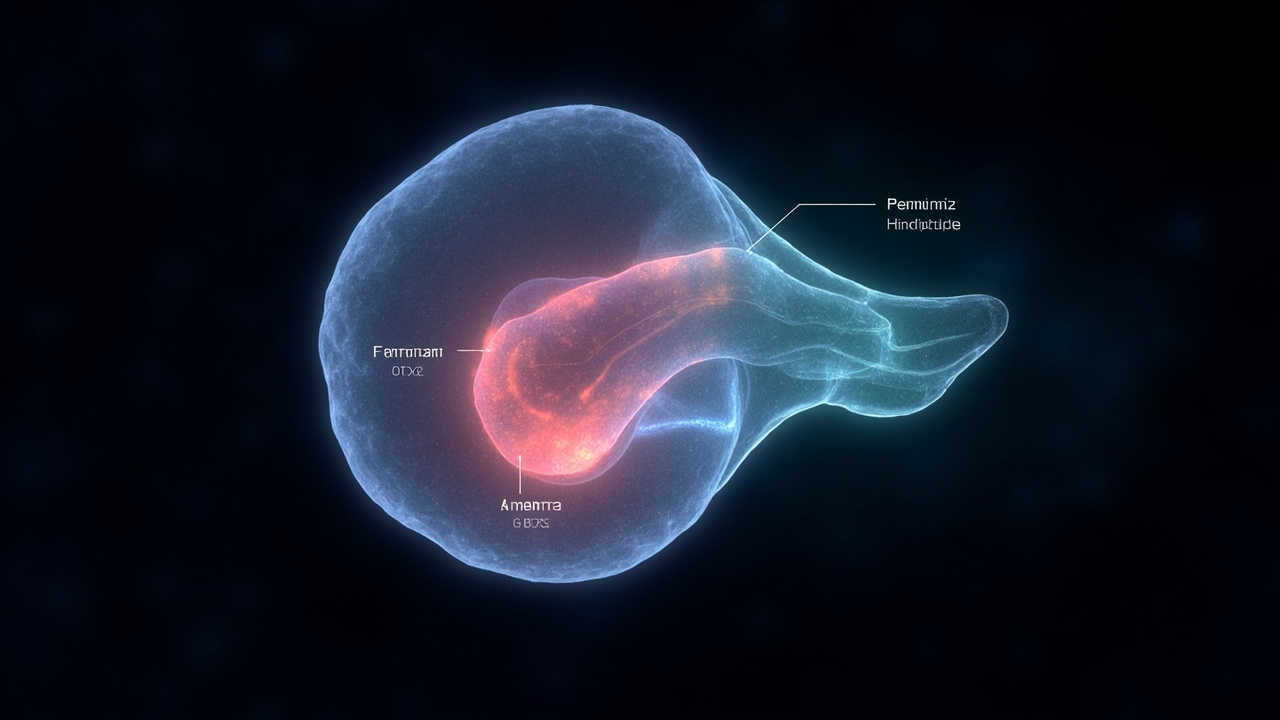
TL;DR: You have one brain, not two — but that single organ starts from two separate starter cell groups that run on parallel tracks from the earliest days of embryonic life.
A Stanford Medicine-led study published Sept. 18 in Nature Neuroscience found that the forebrain and midbrain arise from a different progenitor than the hindbrain, a result that was quickly stretched into headlines about humans having two brains. According to Ars Technica, which examined the paper on Sept. 23, that framing is a silly way to read the findings, because the adult organ is continuous and unified even though its beginnings are split.
The real story, both outlets agree, is about how position is assigned in a developing nervous system, and why laboratory efforts to grow hindbrain cells kept failing for decades.
How "two brains" headlines started
Late last week, news aggregators began carrying claims that scientists had discovered humans may actually have two brains instead of one, according to Ars Technica. That outlet traced the wave to a single press release put out by one of the universities where the work was done.
The underlying research came from Stanford University developmental biologist Kyle Loh and graduate students Carolyn “C” Dundes and Rayyan Jokhai, identified as co-first authors in the Stanford Medicine announcement. The findings were published Sept. 18 in Nature Neuroscience under the title “Two parallel neural ectoderm progenitors contribute to the developing brain,” according to the journal’s listing.
Stanford Medicine’s announcement itself used two-brain language, stating that the human brain is two distinct organs adjacent to one another and that the finding could aid research into diseases affecting the brain stem. In an interview with Nautilus published Sept. 18, Loh said the paper faced an uphill battle to publication because the idea conflicted so strongly with the long-held view that the brain has a common origin, recalling that a department chair once stood up after a conference talk and declared the claim completely false.
Ars Technica science editor John Timmer pushed back on the shorthand. An MRI scan still shows one organ, he noted, and calling the result “two brains” obscures the developmental mechanism the authors actually documented.
One organ, four segments
To understand what was found, it helps to recall how vertebrate nervous systems are divided. According to Ars Technica, vertebrates have four major segments: the forebrain, midbrain, hindbrain, and spinal cord, with further subdivisions forming later inside some of those regions.
Developmental biology studies how a single fertilized egg produces all adult tissues through timed and choreographed steps, the same outlet explained. By the time the first cells committed to becoming neurons appear, the vertebrate embryo already distinguishes head from tail and back from belly.
At that stage the embryo contains three tissues, as described in that report: endoderm, which will line the gut; mesoderm, which will form muscle and bone; and ectoderm, which will go on to form skin. Neural cells begin as a thickening of ectoderm running down the center of the embryo from head to tail. The center sinks while the sides fold upward, forming a deep V shape that eventually meets and pinches off as an oval-shaped neural tube. Every signal carried by the spine and every processed sound, shape and thought depends on descendants of those tube cells.
That general sequence is shared across vertebrates, but timing varies by species. In mice, neural tissue forms nearly simultaneously along the head-to-tail dimension, while in chickens the process starts in the head and moves slowly toward the tail, so brain structures are already forming while future spinal cord cells have not yet adopted a neural identity, according to the same account. Tadpoles build a relatively simple spinal cord that is later expanded and reorganized during the change into frogs.
The positioning problem
Once that tube exists, its cells look largely alike. That raises the central question highlighted by Ars Technica: how do segments of the nervous system form?
Because the embryo already knows head from tail, two simple possibilities present themselves. One is that ectoderm cells already carry positional information, which nerve cells then inherit. The other is that neighboring non-neural cells send signals after neural cells form, with different collections of signaling molecules in head and tail telling the nervous system where to make brain versus spinal cord.
Earlier work had shown that early embryonic ectoderm activates two genes in different territories, one in the front half and one in the back, providing at least crude inherited position. The new study built directly on that observation. The team modified copies of those genes so fluorescent proteins were produced wherever each gene was translated into protein, allowing the fate of each population to be tracked in engineered mice.
That approach revealed two parallel groups rather than one flexible pool, a point emphasized across all three sources.
Otx2 in front, Gbx2 in back
In an interview with Nautilus, Loh described looking at a mouse embryo seven and a half days old, then just a U-shaped clump of cells with no visible head or tail. It was once thought that all cells inside the embryo could form brain, he said. When his team examined expression of two genes, one called Otx2 and the other called Gbx2, they found two different groups instead of one common brain progenitor.
According to the Stanford Medicine announcement, one progenitor expresses Otx2 and is destined to become forebrain and midbrain, while the other expresses Gbx2 and is committed to forming hindbrain. The two populations never overlap and are mutually exclusive from the earliest stages examined.
Loh told Nautilus that people in the 1990s had already observed those genes turning on in different parts of the embryo. His group’s advance was to show that the distinction marks a lineage restriction at the moment cells gain brain identity, rather than a later decision. In his phrasing, as summarized in that interview, a cell putting on its brain hat already declares whether it will make forebrain or midbrain versus hindbrain, instead of remaining naive and able to make any of the three regions.
The team also examined chromatin, the packaging that determines which genes are accessible and which are bundled away. The Stanford Medicine report states that anterior neural ectoderm, the future forebrain and midbrain, and posterior neural ectoderm, the future hindbrain, have fundamentally different chromatin configurations. Those landscapes foreshadowed future identities and effectively locked each progenitor onto its track, like travelers on parallel lines that never cross.
The paper’s abstract, as listed by Nature Neuroscience, draws the same conclusion: two parallel brain progenitors emerge simultaneously during gastrulation, and human stem-cell derivatives of each show diverging chromatin and lineage commitment to forebrain/midbrain versus hindbrain fates. The authors postulate the brain is a composite organ from two lineage-restricted progenitors.
Why hindbrain cells were so hard to grow
One practical payoff involved human pluripotent stem cells, cells able to create any cell in the body. Despite the importance of the hindbrain, scientists had struggled for decades to generate human hindbrain neurons in the laboratory, according to Stanford Medicine.
Armed with the two-progenitor insight, the researchers coaxed human pluripotent cells into functional hindbrain motor neurons. Those lab-grown neurons displayed waves of electrical activity called action potentials and made proteins that identify segments controlling facial and swallowing muscles, the university reported. The Nature Neuroscience abstract specifically notes generation of hindbrain rhombomere 5/6-specific motor neurons that had been difficult to generate in vitro.
That success explained prior frustration. “Previous attempts to make hindbrain neurons likely tried to coax forebrain and midbrain progenitors into hindbrain cells, which our study shows is not possible,” co-first author Jokhai said, as quoted by Stanford Medicine. In the same announcement, Jokhai added that stem-cell biology often focuses on the end cell type such as the neuron, while attention to the earliest embryonic stages revealed the split.
The medical interest centers on the brain stem. The adult brain’s three main regions were described in that announcement as forebrain for higher-level thinking including language, consciousness and abstract reasoning; midbrain as an intermediate zone; and hindbrain at the back of the skull, often called the brain stem, for automatic functions including breathing, sleeping, and regulating heartbeat and hunger urges. Hindbrain neurons also control muscles of the face, tongue and throat involved in speech and swallowing.
Two diseases illustrate the stakes. Spinal muscular atrophy, also known as SMA, is a leading genetic cause of death in children under 1 year of age, while amyotrophic lateral sclerosis, also known as ALS or Lou Gehrig’s disease and often diagnosed between ages 40 and 70, affects both forebrain and hindbrain, according to the same source. In both disorders certain hindbrain neurons gradually cease to function, with loss of swallowing that can cause pneumonia when food or liquid enters the lungs, followed eventually by loss of breathing ability. Reliable hindbrain neurons in a dish would give researchers a new way to study those processes.
Challenging a model from 1951
The result confronts a model dating to 1951 or 1952, when Dutch embryologist Peter Nieuwkoop proposed a common progenitor cell for the entire brain, as recounted by Loh in Nautilus. That idea made intuitive sense because forebrain, midbrain and hindbrain are contiguous in adults, Loh said, comparing the assumption to viewing the lung’s two lobes as obviously from one origin because they sit together.
For decades researchers subscribed to the single-progenitor theory, which held that all parts shared a common developmental origin, according to Stanford Medicine. The new lineage-tracing work in mouse embryos supports the alternative: multiple neural ectoderm progenitors, each restricted to specific regions.
Evolutionary history was part of the argument. The study found the same two-origin pattern in chickens, zebrafish and acorn worms, tiny ocean-floor creatures sharing a distant common ancestor with mammals, according to the university summary. The paper’s abstract places that conservation across 550 million years from hemichordates to mammals.
Loh told Nautilus that perspective changes if one starts with jellyfish, which do not have one brain but have two physically separate nervous systems. Had scientists studied those animals first, he suggested, they might have assumed separate sources pushed together during evolution to create the appearance of one brain, rather than assuming a single origin because mice and humans present one large structure inside a skull.
The literary framing came from physician and author Lewis Thomas, whose 1979 book The Medusa and the Snail won the 1981 National Book Award. Thomas called the transformation of a single cell into a brain capable of building airplanes and writing sonatas one of the great astonishments of the earth, urging endless wonderment, then added in a footnote, “Maybe not one, but two cells,” as quoted in Nautilus. Loh cited that footnote when describing why the new data felt like fulfillment of an old hunch.
Even so, Ars Technica cautioned against turning a developmental split into an anatomical count. The forebrain does the elaborate work of thinking while the hindbrain sustains life-support functions, but both remain parts of one continuous adult brain built from two starting populations. In that reading, humans do not walk around with two brains — they develop one brain from two distinct sets of instructions that meet, merge and cooperate.
Sources referenced
- Contrary to some reports, you don't have two brains - Ars Technica (https://arstechnica.com/science/2026/09/contrary-to-some-reports-you-dont-have-two-brains/)
- Human brain is two separate organs, Stanford Medicine-led research finds (https://med.stanford.edu/news/all-news/2026/09/two-separate-brains.html)